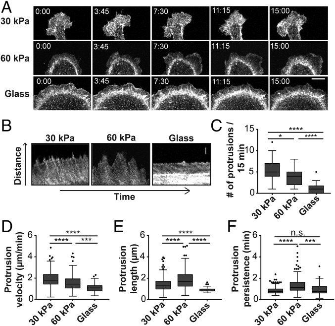
Fig. 2.
Ecad-Fc substrate rigidity influences actin dynamics in adhering cells. (A) Time-lapse images of MDCK cells expressing GFP-LifeAct adhered to Ecad-Fc–functionalized surfaces of varying rigidities. (Scale bar, 10 µm.) (B) Representative kymograph images of membrane protrusions from MDCK cells expressing GFP-LifeAct adhered to Ecad-Fc substrates. (Scale bar, 2 µm.) (C–F) Quantification of number of protrusions (C), protrusion velocity (D), protrusion length (E), and protrusion persistence (F) over a 15-min interval. Four regions from at least 10 different cells were analyzed for each substrate, resulting in quantification of 209 protrusions (30 kPa), 143 protrusions (60 kPa), and 43 protrusions (glass). Results are represented in a box and whisker format, in which the ends of the box mark the upper and lower quartiles, the horizontal line in the box represents the median, and whiskers outside the box extend to the highest and lowest value within the 1.5× interquartile range. Statistics were determined using a Kruskal–Wallis test with Dunn’s posttest for multiple comparisons, *P < 0.05, ***P < 0.0002, ****P < 0.0001.

