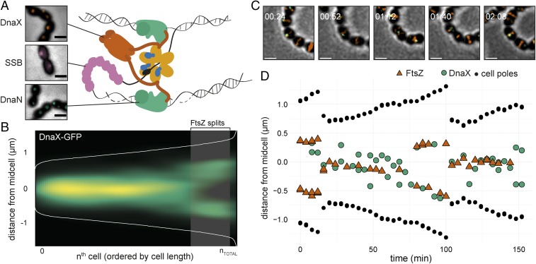
Fig. 2.
Localization of the pneumococcal replisome. (A) Localization of DnaX-GFP (RR31), GFP-DnaN (DJS02), and SSB-GFP (RR33). (Scale bar, 1 μm.) A cartoon of the bacterial replication fork at initiation shows the role of DnaX (clamp loader), SSB, and DnaN (β sliding clamp). DNA polymerase is replicating the leading strand (Top) and Okazaki fragments at the lagging strand (Bottom). The cartoon is based on what is known about the replication fork in E. coli. (B) Plotting the localization of DnaX-GFP (RR22) shows that the replisome is localized at midcell. Data are from a total of 3,574 cells and 3,214 unique localizations. The shaded area represents the point in the cell cycle where 50% of FtsZ has localized to the 1/4 positions of the cell. (C) Snap shots from a representative time-lapse movie of strain MK396 (dnaX-GFP, ftsZ-mKate2). Overlays of GFP, RFP, and phase contrast are shown. (Scale bar, 1 µm.) (D) Transcript of the cell shown in C. The distance of FtsZ (red), DnaX (green), and the cell poles (black) to midcell is plotted against time. The data are also shown in Movie S1. Transcripts of more single cells are shown in SI Appendix, Fig. S3.

