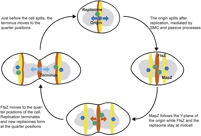
Fig. 6.
A schematic model for division site selection in the pneumococcus. The bulk chromosome is shown in gray, whereas the chromosomal origin, left/right arm, and terminus are indicted as a dark blue hexagon, green circle, and light blue diamond, respectively. MapZ is shown in yellow and FtsZ in orange. Four key stages of the pneumococcal cell cycle, with a newborn cell on the top, are included.

