Fig. 1.
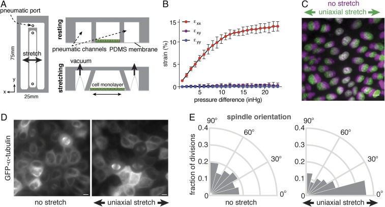
Epithelial cell divisions align with a low level of uniaxial stretch. (A) Design of uniaxial cell stretching device, in which a stretchable silicone membrane is bonded to the bottom of microfabricated PDMS channels. The central channel containing cells is surrounded with pneumatic channels in which vacuum pressure is controlled to induce outward deflection of the channel walls surrounding the cell chamber and stretching of the underlying membrane. (B) Substrate strain measured by displacement of microsphere beads in the normal (εyy), transverse (εxx), and shear (εxy) directions, with the mean percentage of strain and SD from three independent experiments. (C) Representative example of 12% axial cell strain based on the displacement of nuclei in confluent MDCK monolayers, showing the distribution of nuclei before (magenta) and after (green) application of stretch. (D) Visualization of the mitotic spindle in MDCK cells expressing GFP–α-tubulin with and without application of 12% uniaxial stretch (3 h of stretch in this representative image). (E) Circular frequency histograms (Rose diagrams) of the mitotic spindle angle relative to the direction of stretch, in MDCK GFP–α-tubulin cells with (n = 279) or without (n = 153) application of at least 1 h of uniaxial stretch, from three independent experiments. Orientation of cell divisions in the direction of stretch is observed within 1 h of stretch (Fig. S1C). (Scale bars, 10 µm.)

