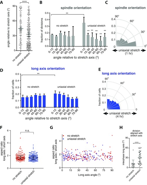
Fig. S1.
A low level of uniaxial stretch orients cell divisions, whereas only having a minimal effect on cell shape. (A) Unbinned data of the mitotic spindle angle relative to the direction of 12% stretch, from three independent experiments (shown as Rose diagrams of binned data in Fig. 1E). Gray bars show the mean spindle angle with SD from the independent experiments. ****P < 0.0001 using a Mann–Whitney U test. (B) Bar graph of the distribution of the division orientation relative to the direction of 12% stretch, showing the mean with SD from three independent experiments (shown as Rose diagrams in Fig. 1E). **P < 0.007 using an unpaired Student t test. (C) Circular frequency histograms (Rose diagrams) of the mitotic spindle angle relative to the direction of stretch, in MDCK GFP–α-tubulin cells with application of 1 h of uniaxial stretch (n = 108 cells), from three independent experiments. (D) Bar graph of the distribution of the cell long axis relative to the direction of 12% stretch, showing the mean with SD from six independent experiments (shown as binned Rose diagrams in Fig. 2B). **P < 0.007 using an unpaired Student t test. (E) Circular frequency histograms (Rose diagrams) of the distribution of the long axis angle relative to the direction of stretch in MDCK E-cadherin:DsRed cells after 4 h of uniaxial stretching, from three independent experiments with at least 500 cells per experiment (for data of long axis orientation of unstretched cells and cells stretched for 2 min, see Fig. 2B). (F and G) Aspect ratio of the long and short axes of MDCK E-cadherin:DsRed cells before and after 12% uniaxial stretch (F) and distribution of the aspect ratio relative to the angle of applied stretch (G). Data are pooled from seven independent experiments; n = 122 unstretched and 160 stretched cells (of which 96 and 120 had an aspect ratio of >1.4, respectively). n.s., not significant, using a Mann–Whitney U test. (H) Angle of the cell long axis (in cells with aspect ratio >1.4) that have their mitotic spindle aligned with uniaxial stretch (within 15°) or in the same direction before application of stretch, showing that cell divisions that are aligned with uniaxial stretch have no strong bias in long axis orientation. Gray bars indicate the mean long axis angle with SD from data from seven independent experiments (15 unstretched and 35 stretched cells). ****P < 0.009.

