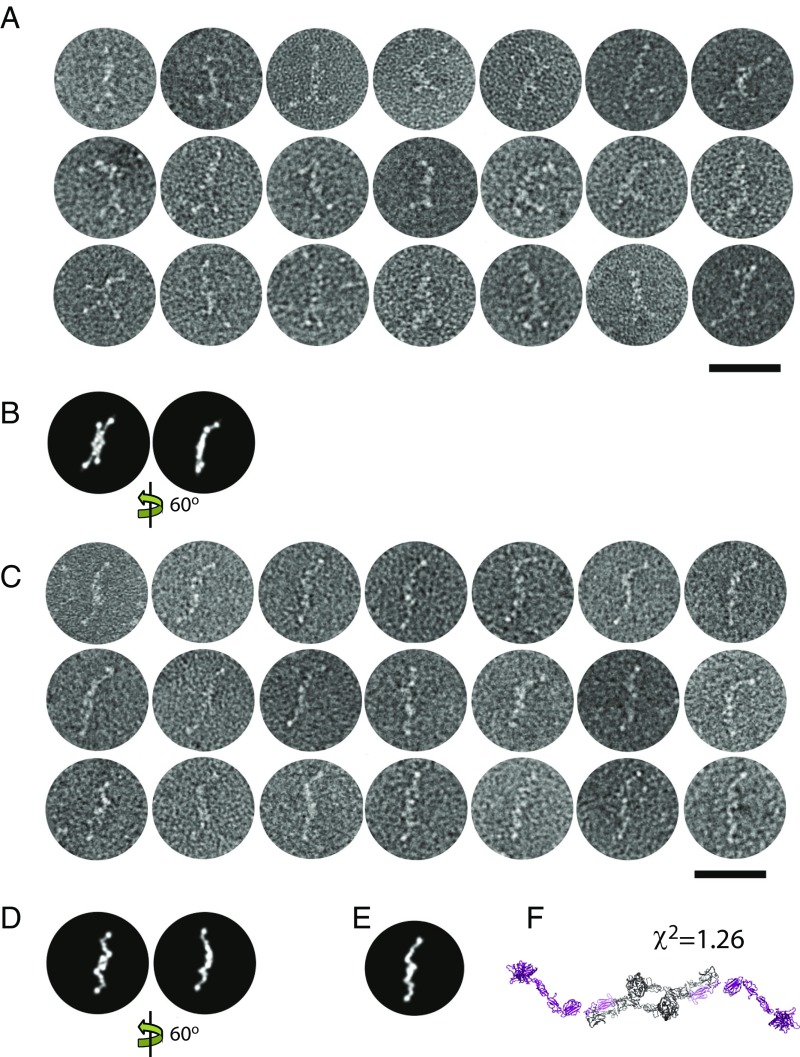
Fig. 2.
Ns EM of selected particles of the free C1r2s2 tetramer. Raw images from the dataset in our study (2) are shown without averaging. (Scale bar: 50 nm.) (A) Particles with split ends. (B) Two computed projection images of the SAXS model our study (2). (C) Particles with simple ends. (D) Projection images of the SAXS model shown in Fig. 1. (E and F) Projection image of a SAXS model with more extended C1s domains compared with the SAXS model in Fig 1. Note that the SAXS models in both figures are symmetrized twofold.

