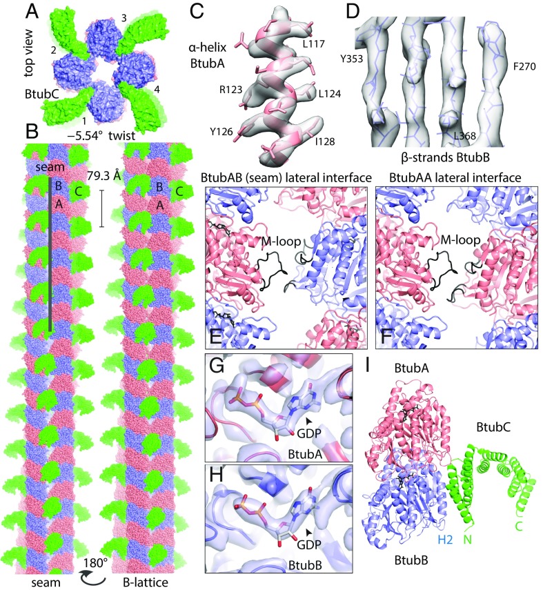
Fig. 2.
BtubABC mini microtubule cryoEM structure at 3.6 Å resolution. (A) View along the filament axis showing the four protofilaments end-on and also the protruding BtubC subunits in green. (B, Left) BtubABC filaments. PdBtubA is in red, PdBtubB in blue, and PvBtubC in green. The view shown highlights the seam (A lattice, A-B lateral contacts). (B, Right) Rotated by 180°, showing the B lattice (A-A and B-B lateral contacts). Helical parameters: twist = –5.54°, rise = 79.3 Å. (C and D) CryoEM density map with fitted atomic model. (E) Close-up of the only lateral contact holding the protofilaments together. The contact is related to the equivalent contact in eukaryotic microtubules, with the M loop (3) of BtubA (residues ∼280–290) reaching over to contact residues around 90 and also 55–65. Shown here is the seam contact (BtubA M loop reaching over to BtubB). Note that the M loop in BtubB is not ordered. (F) Same as in E, but B-lattice contact, BtubA contacting BtubA. (G) Close-up of density in the nucleotide binding pocket in BtubA, which is best fitted with GDP. (H) Pocket in BtubB, also GDP-bound. (I) Cartoon plot of the interaction of BtubC with the BtubAB heterodimer. BtubC predominantly binds to BtubB. See also Movie S3.

