Fig. 2.
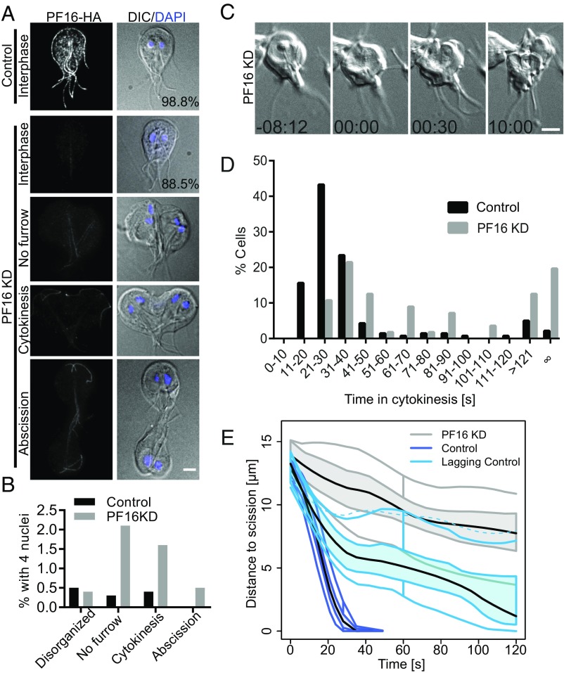
Flagella function is required for furrowing progression and abscission. (A) PF16 localization in control and PF16-KD cells with equal exposure and scaling. Note that 98.8% of control and 88.5% of PF16-KD cells are normally organized interphase cells. (B) Categorization of the 1.2% of control and 11.5% of PF16 KD cells with four nuclei. With the exception of disorganized cells, which lacked normal polarity and could not be scored, images representing the categories are shown in A. (C) Still images from a time-lapse movie of a PF16-KD cell that failed to complete cytokinesis. (D) Histogram of division times for PF16-KD cells (n = 56) and morpholino control cells (n = 141). Data acquired from at least three independent replicates. (E) Functional box plot of furrow trajectories for the first 2 min of 11 PF16-KD cells that never completed cytokinesis (gray), compared with 20 randomly sampled control cells (blue) and the 11 slowest control cells (light blue). Central black line is the mean of the bootstrapped LOESS curves; the colored band indicates the 50% CI bound by 95% confidence bands of bootstrapped distances.

