Fig. 3.
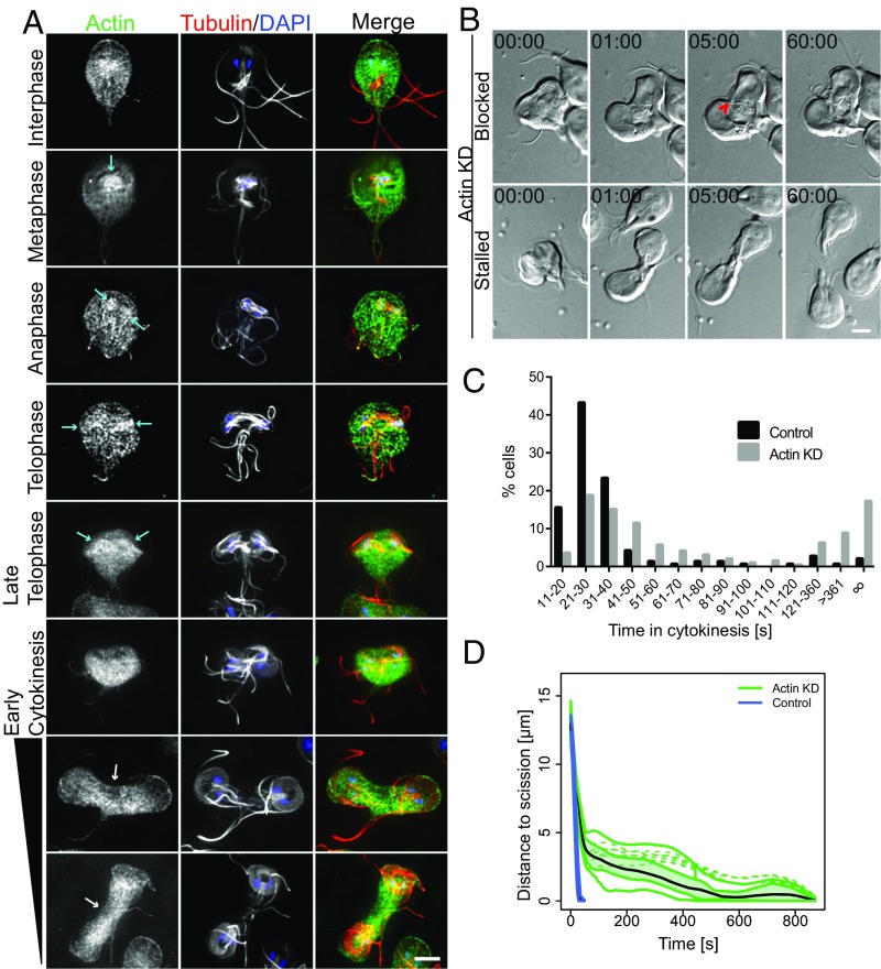
Actin is required for abscission. (A) Immunofluorescence localization of Actin (green), tubulin (red), and DNA (blue) throughout the cell cycle. Actin accumulated around spindles and developed daughter ventral discs (cyan arrows). During cytokinesis, actin is found at relatively lower levels at the leading edge of the cleavage furrow (white arrows; analysis in Fig. S5). (B) Live cell imaging of actin KD reveals blocked and stalled cell phenotypes. Block of cytokinesis resulted from a flagella crossing the cleavage furrow path (red arrowhead; Movie S5). The stalled cell completes cytokinesis after ∼60 min (Movie S6; time stamp, min:s). (Scale bars: 5 μm.) (C) Histogram of time to complete cytokinesis. Actin-KD cells (n = 191) take longer to divide than cells treated with the morpholino control (n =141). Data acquired from at least three independent experiments. (D) Functional box plot of bootstrapped LOESS curves for actin-KD cells (green, n = 12) taking 6–15 min to divide compared with typical control cells (blue, n = 20) shows a delay in abscission.

