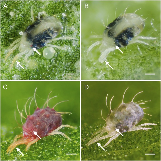
Fig. 2.
The albino pigment phenotypes of T. urticae and P. citri. Shown are (A) WT T. urticae pigmentation (strains Wasatch, MAR-AB, Foothills, and London; a representative London mite is shown), (B) albino phenotype of T. urticae (strains Alb-NL, Alb-JP, W-Alb-7, -8, -10, -11, and -14; a representative Alb-NL mite is shown), (C) WT P. citri phenotype, and (D) albino phenotype of P. citri. In all cases, adult females are shown. Arrows indicate red eye spots or red-orange in the distal front legs of WT mites that are absent in the albino mutants. The dark regions present in the mite bodies are gut contents that are visible because spider mites are partly translucent. (Scale bars: 0.1 mm.)

