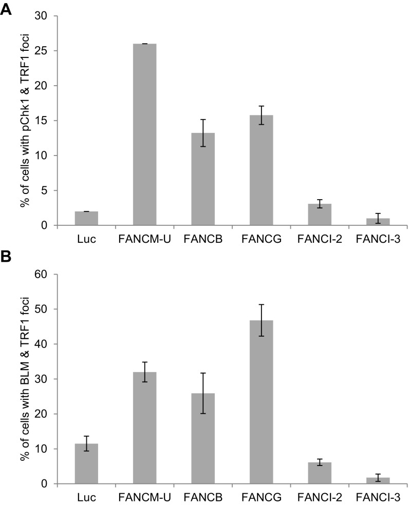
Fig. S7.
The effect of different FA proteins on the Chk1-pS345 and BLM foci formation. U2-OS cells were transfected with siRNA as indicated. Cells were costained with an antibody recognizing TRF1 together with an antibody recognizing Chk1-pS345 (A) or BLM (B). All nuclei were stained with DAPI. More than 200 cells were counted for each sample. All error bars are SD obtained from three different experiments.

