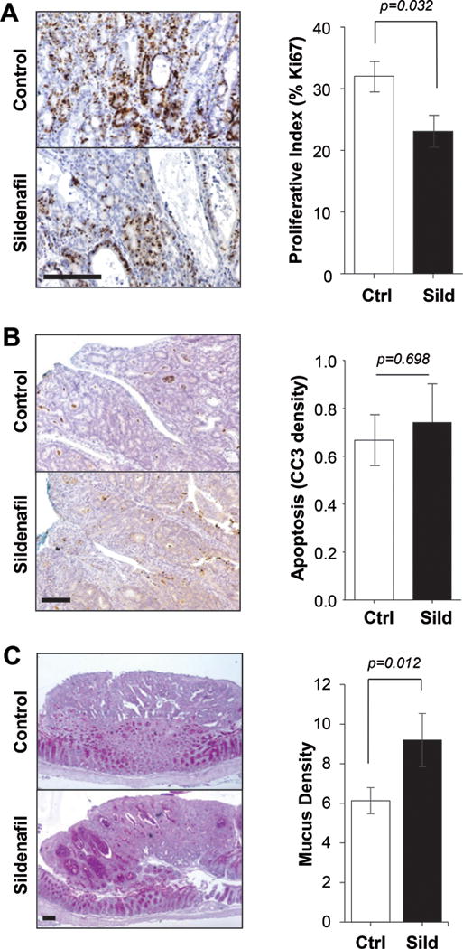
Figure 4. Polyps from sildenafil treated mice are less proliferative and more differentiated.

A, IHC staining of Ki67 (left panel) and quantitation (right panel) in polyps of untreated (Ctrl) and treated (Sild) mice. B, IHC staining of cleaved caspase 3 (CC3) (left panel) and quantitation (right panel) in polyps of untreated (Ctrl) and treated (Sild) mice. C, AB/PAS staining of mucus (left panel) and quantitation (right panel) in polyps of untreated (Ctrl) and treated (Sild) mice. In all panels, the scale bar is 100 μm and n=10 mice per group.
