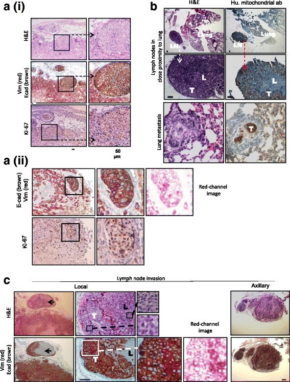
Fig. 2.

Pronounced expression of E-cadherin was observed in MDA-MB-468 xenograft metastases. a (i) and (ii) Similarity of features of MDA-MB-468 xenograft tumors. Red-channel image is shown for simplicity of vimentin staining. b Metastatic tumor cells in lymph nodes (LNs) and lungs. Positive staining for an antihuman mitochondrial antibody confirmed that tumor cells were of human origin (T), whereas mouse lymphocytes (L) were not stained. Tumor cells in the lung demonstrated E-cadherin expression but not vimentin. c LN invasion. Black arrows indicate invasion of E-cadherin and vimentin-expressing tumor cells to the adjacent LNs (T). Red-channel image is shown for clarity of vimentin staining. Tumor cells in axillary nodes stained homogeneously for E-cadherin. Lymphoid tissue (L) did not stain for either E-cadherin or vimentin, confirming its murine origin. All scale bars = 50 μm, except for axillary LN images, where scale bar represents 100 μm. H&E Hematoxylin and eosin
