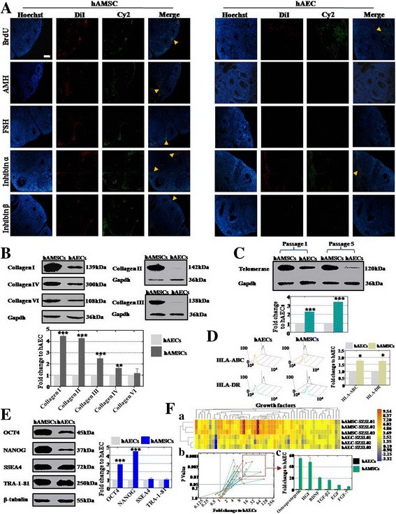
Fig. 6.

Distinction of cellular biological characteristics between hAMSCs and hAECs. a Expression of ovarian markers (AMH, FSH, inhibin α, and inhibin β) and proliferation marker (BrdU) in ovarian tissue measured after hAEC and hAMSC transplantation respectively. b Secretory level of collagen (I, II, III, and IV) from hAECs and hAMSCs estimated by western blot analysis respectively. c Activity of telomerase in hAECs and hAMSCs tested by western blot assay at passage 1 and 5 respectively. d Expression level of HLA-ABC and HLA-DR in hAECs and hAMSCs tested by FACS respectively. e Expression level of pluripotency markers (OCT4, NANOG, SSEA4, and TRA-1-81) in hAECs and hAMSCs measured by western blot analysis. f Growth factor derived from hAECs and hAMSCs estimated by antibody microarray respectively (a–c). fa heatmap exhibited the secretory level of growth facors between hAMSCs and hAECs; fb distribution of 52 growth factors were demonstrated after secretory level of hAMSCs compared to hAECs; fc in accordance with standard criteria of fold change ≥ 8 and statistical significance (p < 0.01), six growth factors were selected: osteoprotegerin, HGF, BDNF, TGF-β2, EGF, and FGF-7. All experiments were carried three times. Error bars indicate SD. *p < 0.05, **p < 0.01, ***p < 0.001, compared with hAECs group. hAEC human amniotic epithelial cell, hAMSC human amniotic mesenchymal stem cell
