
Fig. 3.
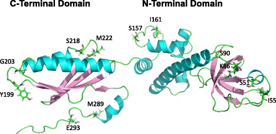
Spatial distribution of structurally comparable residue pairs from eIF2α. Ribbon cartoon of the three-dimensional structure of the α subunit of human eIF2 (PDB 1Q8K). The modifiable residues (either Ser, Thr, Tyr or Met) located outside helices and strands are displayed using stick representation, as well as those amino acids found four positions away from them