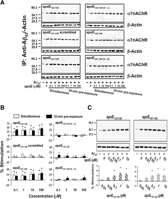
Fig. 1.

ApoE141–148 mediates apoE-induced Aβ42-α7nAChR association enhancement ex vivo in rat brain synaptosomes. Rat frontal cortical synaptosomes were incubated with 0.1–100 μM apoE either 10 min prior to or simultaneously with 0.1 μM Aβ42. Synaptosomes were collected by centrifugation, solubilized, and immunoprecipitated with anti-Aβ42. The level of Aβ42-associated α7nAChRs in anti-Aβ42 antibody immunoprecipitates was shown by Western blot detection of α7nAChR a and quantified by densitometric scanning (b). Separately, rat cortical synaptosomes were incubated with 0.01, 0.05, 0.1, 1, and 10 nM of apoE133–149 or apoE141–148 simultaneously with 0.1 μM Aβ42. The level of Aβ42-associated α7nAChRs in anti-Aβ42 antibody immunoprecipitates was demonstrated by Western blot detection of α7nAChR and quantified by densitometric scanning (c). *p < 0.01, compared to Aβ42 alone by Newman-Keuls multiple comparisons (n = 5). α7nAChR α7-nicotinic acetylcholine receptor, Aβ amyloid beta, ApoE apolipoprotein E, IP immunoprecipitation
