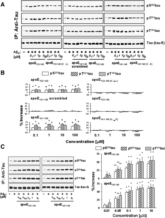
Fig. 5.

ApoE141–148 mediates ApoE-induced Aβ42-elicited α7nAChR-dependent tau phosphorylation. Rat frontal cortical synaptosomes were incubated simultaneously with 0.1–100 μM apoE fragments and 0.1 μM Aβ42. Synaptosomes were collected by centrifugation, solubilized, and immunoprecipitated with anti-tau antibodies. The levels of Aβ42-induced tau phosphorylation on the serine 202 (pS 202 tau), threonine181 (pT 181 tau), and threonine231 (pT 231 tau) in anti-tau immunoprecipitates shown were determined using Western blot detection of each phosphoepitope a and quantified by densitometric scanning (b). Separately, rat cortical synaptosomes were incubated with 0.01, 0.05, 0.1, 1, and 10 nM apoE133–149 or apoE141–148 simultaneously with 0.1 μM Aβ42. The level of Aβ42-induced pS202tau, pT181tau, and pT231tau in anti-tau immunoprecipitates were determined by Western blot detection of each phosphoepitope and quantified by densitometric scanning (c). *p < 0.01, **p < 0.05, compared to Aβ42 alone by Newman-Keuls multiple comparisons (n = 4–6). Aβ amyloid beta, ApoE apolipoprotein E, IP immunoprecipitation
