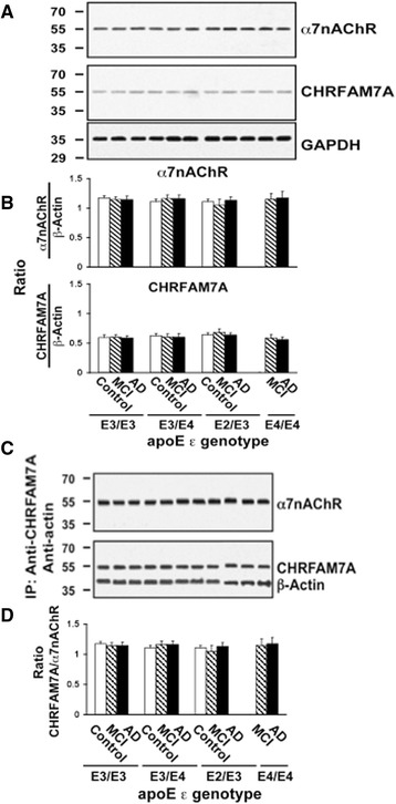
Fig. 8.

No APOE genotype- or diagnosis-related changes in α7nAChR and CHRFAM7A expression levels in lymphocytes. Lymphocytes obtained from cognitive normal controls, subjects with mild cognitive impairments (MCI) and Alzheimer’s disease (AD) were solubilized. The expression levels of α7nAChR and CHRFAM7A, both with apparent molecular mass of 54 kDa, in 50 μg of solubilized lymphocytes along with the loading control, GADPH, are shown by Western blot detection a and quantified by densitometric scanning that demonstrates no discernible changes in α7nAChR or CHRFAM7A expression (b). Solubilized lymphocyte membranes (200 μg) were used to assess α7nAChR/CHRFAM7A complex levels by immunoprecipitation with immobilized anti-CHRFAM7A and -actin. The abundance of α7nAChR, CHRFAM7A, and β-actin in anti-CHRFAM7A/actin immunoprecipitate is shown by Western blot detection c and quantified by densitometric scanning that demonstrates no diagnosis- or APOE ε genotype-related changes in α7nAChR, ChRFAM7A, and β-actin levels in lymphocyte membranes (d). n = 86 including 32 AD, 30 MCI and 24 control subjects from four different APOE genotype groups. α7nAChR α7-nicotinic acetylcholine receptor, ApoE apolipoprotein E, IP immunoprecipitation
