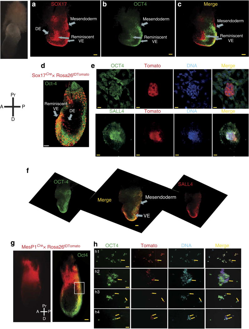
Figure 1. Embryonic pattern of expression of SOX-17/OCT4/SALL4.
Whole-mount staining of E7.5 mouse embryos with (a) anti-SOX-17- and alexa546-conjugated secondary antibody, (b) anti-OCT4- and alexa488-conjugated secondary antibody, (c) merged image (a,b). (d) Anti-OCT4 whole-mount immunostaining of E7.5 embryos derived from a Sox17cre/+ breeder crossed with Rosa26tDTomato female. (e) the embryo proper was enzymatically dissociated and the cells were plated and fixed on coverslip. The cells were immunostained by an anti-OCT4 (upper panel) or anti-SALL4 antibody. Scale bars, 10 µm. Note that OCT4 is cytosolic in most of cells indicating a downregulation of the transcription factor. (f) Whole-mount staining of embryo using an anti-OCT4 and anti-SALL4 antibodies; a stack of images was acquired in confocal microscopy and reconstituted as a 3D image using AMIRA software. The merged image is shown in the centre. DE, definitive endoderm, VE visceral endoderm. (g) Anti-OCT4 immunostaining of E7.5 embryos derived from a MesP1cre/+ breeder crossed with Rosa26tDTomato female. Whole-mount staining. (h) The posterior region of the embryo was enzymatically dissociated and cells were plated and immediately fixed on coverslip to be stained by an anti-OCT4 antibody and a secondary anti-alexa488 secondary antibody. Arrows indicate OCT4 + cells in the MesP1 cell lineage. OCT4 is either nuclear (h1) or cytosolic (h2, h3 and h4). Scale bars, 50 µm.

