
Fig. 5.
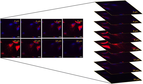
Internalization of HLSC-EVs in hepatocytes differentiated from ASS1-HLSCs. Representative confocal microscopy showing the internalization of 1 × 10¹⁰ Dil-labeled EVs in hepatocytes differentiated from ASS1-HLSCs after 6 h of incubation at 37 °C. Z stack analysis shows the presence of EVs within the cytoplasm of the cells indicating an effective uptake of vesicles; scale bar = 50 μm. Data represent one of three experiments performed with similar results