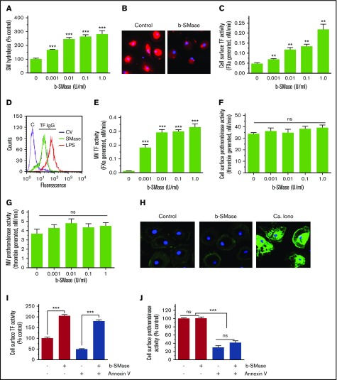
Figure 2.
SM hydrolysis in the outer leaflet of plasma membrane increases cell surface TF activity and releases TF+MVs in macrophages. (A) Human MDMs were metabolically labeled with [methyl-14C]-choline chloride (0.2 μCi/mL) for 48 hours. The labeled cells were treated with a control vehicle or varying concentrations of b-SMase for 60 minutes, and supernatants were collected. The cell supernatants were subjected to centrifugation at 400g for 5 minutes and then 21 000g for 60 minutes to remove cell debris and pellet MVs, respectively. The supernatants devoid of cell debris and MVs were counted for the radioactivity to determine the release of [14C]-phosphorylcholine. (B) MDMs treated with a control vehicle or b-SMase (1 U/mL) for 60 minutes were fixed and incubated with lysenin (0.5 µg/mL), a specific binding protein to SM, for 1 hour. The bound lysenin was detected by using anti-lysenin antiserum followed by secondary antibodies conjugated with AF567 fluorophore. (C) MDMs were treated with varying concentrations of b-SMase for 1 hour. At the end of the treatment, cell supernatants were removed, cells were washed, and cell surface TF activity was measured by adding 10 nM FVIIa and 175 nM FX and determining the rate of FXa generation in a chromogenic assay. (D) MDMs were treated with a control vehicle or b-SMase (1 U/mL) for 1 hour. Intact, nonpermeabilized cells were labeled with control IgG (“C”) or rabbit anti-human TF IgG, followed by fluorophore-conjugated secondary antibodies. The immunostained cells were subjected to FACS analysis. As a positive control, MDMs were stimulated with 1 µg/mL lipopolysaccharides from Escherichia coli O111:B4 (LPS) for 4 hours to enhance TF antigen levels on the cell surface by inducing de novo synthesis of TF. (E) MDMs were treated with varying concentrations of b-SMase for 1 hour. At the end of the treatment, cell supernatants were removed, and MVs were isolated as described in “Materials and methods.” TF activity in MVs was measured as the rate of FXa generated by adding 10 nM FVIIa and 175 nM FX. (F) Cell surface prothrombinase activity of MDMs treated with a control vehicle or varying concentrations of b-SMase for 1 hour was determined by incubating MDMs with FVa (10 nM), FXa (1 nM), and prothrombin (1. 4 µM) and measuring the amount of thrombin generated in a chromogenic assay. (G) Prothrombinase activity in MVs harvested from MDM treated with varying concentrations of b-SMase for 1 hour. The reagent concentrations used for the assay were same as in panel F. (H) AF488–annexin V labeling of MDMs treated with a control vehicle or b-SMase (1 U/mL) for 1 hour. As a positive control, MDMs were treated with calcium ionophore (10 µM) for 10 minutes. Effect of annexin V on cell surface TF (I) and prothrombinase (J) activities. MDMs were pretreated with annexin V (400 nM) for 30 minutes and then treated with a control vehicle or b-SMase (1 U/mL) for 1 hour. *P < .05; **P < .01; ***P < .001 compared with the values obtained in the absence of b-SMase treatment.

