
Figure 4. Antigen density directly impacts T cell activation by B2-OKT3 and huNKG2D-OKT3 BiTEs.
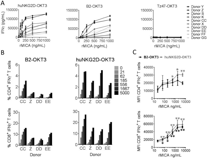
A) Activated T cells from ten donors were incubated with immobilized rMICA at the indicated concentrations and either huNKG2D-OKT3 (left), B2-OKT3 (center) or negative control BiTE (right) at 250 ng/mL for 24 hours. IFNγ from cell free supernatant was measured by ELISA. (B) Cells from 2-4 donors were treated as in A, but after a six hour incubation in the presence of brefeldin A, cells were stained for expression of CD4, CD8 and intracellular IFNγ, and data were acquired by flow cytometry. Percentage of activated cells as indicated by IFNγ+ staining are shown in B, while MFI for each T cell subset over the range of antigen densities tested is shown in C. MFI were compared to 0 ng/ml group by paired t test (* p ≤ 0.05, **p ≤ 0.01). Error bars represent SD of triplicates in (A) and between donors in (C). Blood donors are designated by letters (K, S, P, X, Y, Z, CC, DD, EE, FF, GG).