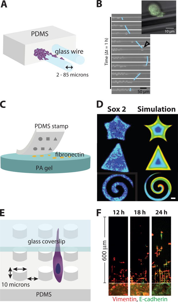
Fig. 3.
2D Cell Monolayers in Microfabricated Geometries. (A) Microscale glass wires embedded in PDMS, (B) Individual cells can detach and scatter along glass wires from multicellular groups. Reproduced from87 with permission from the National Academy of Sciences. (C) Microcontact printing using PDMS stamps to pattern fibronectin shapes on soft PA gels. (D) Cell monolayers display stem-like markers at the periphery and corners of these shapes. Reproduced from88 with permission from Nature Publishing Group. (E) Cells undergoing confined migration within arrays of microscale PDMS pillars, with a glass ceiling. (F) Cells transition from collective to individual migration after EMT. Reproduced from89 with permission from Nature Publishing Group.

