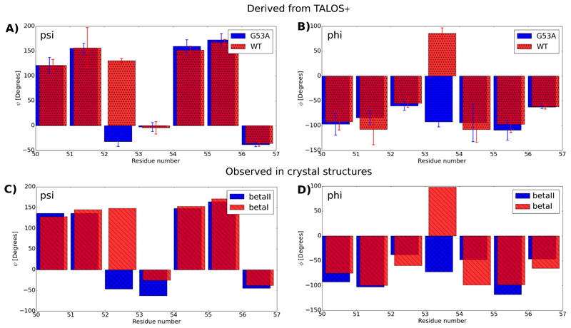
Figure 4.
G53A in MPD-ub crystals forms predominantly a type-I β-turn. (a) and (b) show psi and phi backbone dihedral angles in WT MPD-ub and G53A MPD-ub, as derived from the assigned chemical shifts, and the program TALOS+[43]. (c) and (d) show the corresponding dihedrals in crystal structures forming a type-I β-turn (here the structure 4XOL[48] was used, which is one out of many βI-forming PDB entries, shown in blue) and type-II β-turn (red, PDB entry 3ONS [32]). The comparison of the psi (residue 52) and phi (residue 53) angle shows that this peptide plane is rotated by the G53A mutation.

