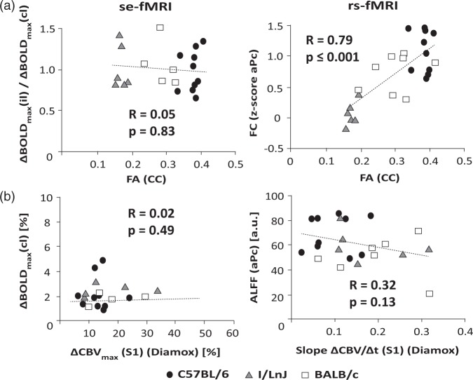
Figure 5.
Correlation of functional readouts from se- and rs-fMRI experiments with structural connectivity and cerebrovascular parameters for individual C57BL/6 (N = 10), I/LnJ (N = 5), and BALB/c (N = 7) mice. (a) Correlation of interhemispheric functional interaction with structural connectivity (FA (CC)). In se-fMRI, interhemispheric interaction is expressed by the ratio of the maximum amplitude of the BOLD response in the contra- and ipsilateral S1HL cortex (ΔBOLDmax(il)/ΔBOLDmax(cl)) following electrical hindpaw stimulation, while in rs-fMRI, it is given by the z-score for the FC in aPc (S1). Analysis reveals a strong positive correlation for FC and FA (R = 0.79) under resting-state conditions; however, no influence of a missing CC was found reflected by the correlation of ΔBOLDmax(il)/ΔBOLDmax(cl) versus FA (R = 0.05). (b) Correlation of local fMRI signal amplitudes, i.e. ΔBOLDmax(cl) derived from the se-fMRI and ALFF in aPc (S1) of the rs-fMRI signal with vascular parameters extracted from the CBV response in S1 elicited by acetazolamide (Diamox) reflecting the vascular reactivity (slope ΔCBV/Δt) and reserve (ΔCBVmax). Neither ΔBOLDmax(cl) nor ALFF in aPc (S1) measured in rs-fMRI showed any dependence on vascular reserve or vascular reactivity, respectively.

