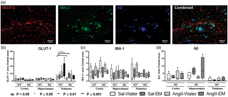
Figure 6.
Distribution of glucose transporter-1 (GLUT-1), ionised calcium-binding adapter molecule 1 (IBA-1), and amyloid-β (Aβ) in the cortex, hippocampus, and thalamus of 12-month-old Sal-infused (saline) and AngII-infused (Angiotensin II) AβPP/PS1 (Alzheimer’s disease, AD) and wild-type (WT) mice, treated with eprosartan mesylate (EM) vs. normal drinking water as control. A representative example of vessel density (GLUT-1), neuroinflammation (IBA-1), Aβ staining, and an overlay of all three staining in an AβPP/PS1 mouse is shown (a). (b) The brains of all mice underwent immunohistochemical staining with GLUT-1 antibody representing amount of GLUT-1. While in the cortical and hippocampal regions, no effect was detected, all AngII-infused animals displayed an increased amount of GLUT-1 in the thalamus compared to their Sal-infused littermates (p = 0.039). Only in thalamic regions of all Sal-infused mice, EM treatment increased amount of GLUT-1 (p = 0.032). (c) The brains of all mice were immunohistochemically stained with an antibody against IBA-1 being specifically expressed in activated microglia. Notably, Aβ and microglia are colocalised as demonstrated in the combined picture. No significant differences were found for IBA-1. (d) The brains of all AβPP/PS1 mice were immunohistochemically stained with WO-2/Aβ antibody (mouse anti-human Aβ4–10). No significant AngII- or antihypertensive treatment effects were found.

