Figure 2.
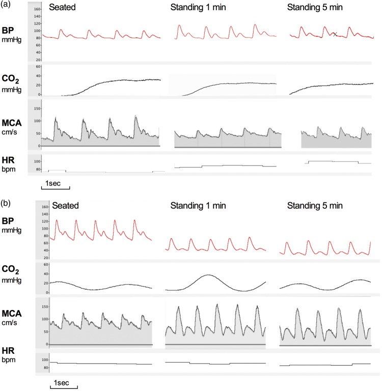
Typical examples continuous blood pressure and MCA velocity. (a) Top panel shows tracings of beat-to-beat blood pressure, heart rate, and MCA velocity with accompanying end-tidal CO2 measurements in a 22-year-old healthy female control sitting and standing. (b) Bottom panel shows the same measurements obtained in a 20-year-old female patient with familial dysautonomia. Note the severe progressive orthostatic blood pressure fall, without compensatory tachycardia in the patient. Despite blood pressure being 60/30 mmHg, she did not complain of symptoms of cerebral hypoperfusion. Systolic and mean MCA flow velocity remained well preserved. The MCA velocity waveform shows increased pulsatility and deeper dichrotic notch, indicating low resistance in the cerebral arterioles to compensate for the decline in stroke volume. BP: blood pressure; CO2: end-tidal CO2 levels; MCA: middle cerebral artery; HR: heart rate. Images were obtained from raw tracings recorded with PowerLab and processed with LabChart 7 (AD Instruments, Dunedin, New Zealand).

