
Figure 4.
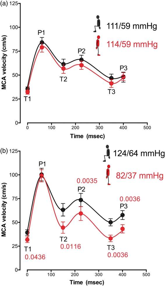
MCA velocity. Key inflection points were identified at three peaks (P1: systolic peak; P2: reflected peak; and P3: following peak) and three troughs (T1: systolic velocity; T2: following trough; and T3: dichrotic notch). Data are averaged for 15 controls (upper panel, (a)) and 25 FD patients (lower panel, (b)), sitting (black lines), and standing (red lines). Average brachial blood pressure sitting (black) and standing (red) at the time of analysis are included. When perfusion pressure was low on standing in patients with FD, systolic flow was maintained while diastolic flow was reduced (panel (b)). Note also the deepening of the dichrotic notch in patients with FD when standing with hypotension. MCA: middle cerebral artery; MCA: middle cerebral artery blood flow velocity; BP: blood pressure. Data are mean ± s.e.m.