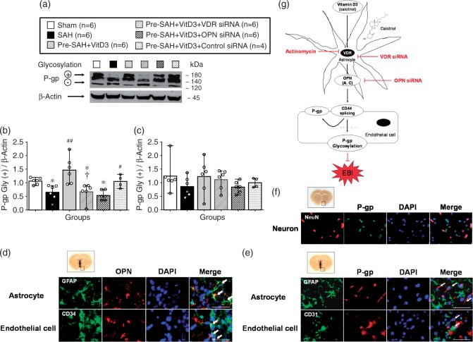
Figure 7.
Vitamin D3 (VitD3) induces P-gp glycosylation after subarachnoid hemorrhage (SAH). Representative Western blots of P-glycoprotein (P-gp) (a) and quantitative analysis of mature/fully glycosylated P-gp (b) and immature P-gp (c) 24 h after SAH. Pre-SAH + VitD3, intranasal treatment 30 ng/kg/day VitD3 24 hours before SAH; siRNA, small interfering ribonucleic acid; *P < 0.05 vs. sham; #P < 0.05 and ##P < 0.001 vs. SAH; †P < 0.05 vs. Pre-SAH + VitD3 + Control siRNA. Colocalization of OPN (red) with astrocytes (GFAP, green) and endothelial cells (CD34, green) (d), and colocalization of P-gp (red) with endothelial cells (CD34, green), but not astrocytes (GFAP, green) (e) and neurons NeuN, red) (f). Nuclei are stained with DAPI (blue). Top panel indicates the location of immunohistochemical staining (small black box). Arrows indicate colocalization. n = 3 per group. Bar = 50 µm. (g) Proposed mechanism of VitD3 neuroprotection via endogenous OPN/CD44/P-gp glycosylation after SAH. EBI, early brain injury.

