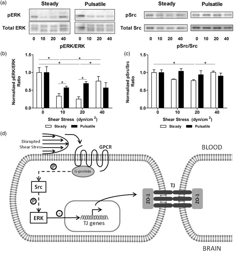
Figure 6.
Effect of shear stress on the activation of Src and ERK1/2. (a) HBMEC were exposed to shear stress for 96 h. Protein was extracted and signaling markers were analyzed with Western blotting. Densitometric analysis of the phosphorylation ratios of pSrc (b) and pERK1/2 (c) showed a minimal activation between 10 and 20 dyn/cm2 and steady flow. At 40 dyn/cm2, phosphorylation increased suggesting that signaling cascades were activated. Similarly, pulsatile flow lead to higher activation profiles. *p ≤ 0.05 between indicated bars. (d) Shear stress disruptions at the brain microvasculature are sensed by G-coupled protein receptors (GPCR). This leads to activation of tyrosine-protein kinases (Src) and extracellular signal-regulated kinases (ERK1/2), which directly induces downregulation of the expression of tight junction markers.
ERK: extracellular signal-regulated kinases; GPCR: G-coupled protein receptor; P: phosphorylation; Src: proto-oncogene tyrosine-protein kinase; TJ: tight junction; ZO-1: Zona Occludens 1.

