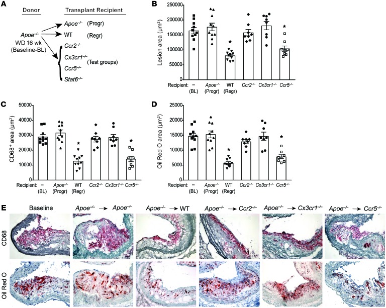
Figure 1. CCR2 and CX3CR1 are required for plaque regression.
Analysis of aortic arch plaques from Apoe–/– mice on 16-week WD (baseline [BL]; n = 10) or 5 days after transplantation into Apoe–/– (progression [Progr]; n = 10), WT (regression [Regr]; n = 10), or chemokine receptor–KO recipient mice (Ccr2–/–, Cx3cr1–/–, or Ccr5–/–; n = 8). (A) Schematic of transplant experiments. Quantification of (B) lesion area, (C) immunohistochemical staining for the macrophage marker CD68, and (D) Oil Red O staining of neutral lipids. *P < 0.001 compared with BL and Apoe–/– progression groups using 1-way ANOVA with Dunnett’s multiple comparisons testing. (E) Representative sections of aortic transplant segments from BL and the recipient groups stained with anti-CD68 antibody and Oil Red O, imaged at ×40 magnification.

