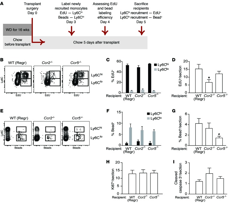
Figure 4. Macrophage dynamics show Ly6chi monocyte recruitment is the key kinetic change impairing regression in Ccr2–/– recipient mice.
Aortic arches from Apoe–/– donors fed WD for 14 weeks were transplanted into recipients. (A) Schematic of timeline for EdU and bead injections into recipient mice to assess recruitment of Ly6Chi and Ly6Clo monocytes, respectively, into transplanted aortic arches under regression conditions. (B) Representative flow cytometry plots of CD45+CD115+ circulating monocytes showing Ly6C versus EdU, and (C) quantification of EdU incorporation in circulating Ly6Chi versus Ly6Clo populations showing that EdU is preferentially incorporated into Ly6Chi monocytes in WT, Ccr2–/–, and Ccr5–/– mice (n = 4–8 per group). (D) Analysis of EdU+ cells/section of atherosclerotic plaques transplanted into WT, Ccr2–/–, or Ccr5–/– recipients showing significantly reduced recruitment into plaques of Ly6ChiEdU+ cells into Ccr2–/– compared with WT recipients. (E) Representative flow cytometry plots of CD45+CD115+ circulating monocytes showing Ly6C versus beads, and (F) quantification of bead incorporation in circulating Ly6Chi versus Ly6Clo monocyte populations showing that beads are preferentially incorporated into Ly6Clo monocytes in WT, Ccr2–/–, and Ccr5–/– mice (n = 8–10 per group). (G) Analysis of bead+ cells/section of atherosclerotic plaques transplanted into WT, Ccr2–/–, or Ccr5–/– recipients showing significantly reduced recruitment of Ly6Clo bead+ cells into Ccr5–/– compared with WT mice. Quantification in plaque sections of (H) Ki67 (to assess proliferation) and (I) cleaved caspase 3 (to assess apoptosis) showed no significant differences in proliferation or apoptosis between transplant recipient groups. Quantifications in D, G, H, and I were done in aortic arch plaques from mice 5 days after transplantation (n = 8–9 per group). #P = 0.05, *P < 0.05 when compared with WT group using 1-way ANOVA with Dunnett’s multiple comparisons testing.

