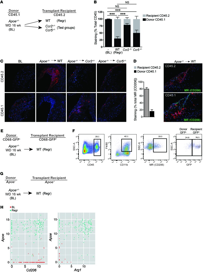
Figure 5. CCR2 deficiency in recipient leukocytes impairs their recruitment to atherosclerotic plaques, where they normally display M2 characteristics.
(A) Schematic of CD45.1 (donor) to CD45.2 (recipient) aortic transplantation experiments with (B) quantification of immunohistochemical staining of CD45.1 and CD45.2 in aortic arch plaques from Apoe–/– mice on 14-week WD (BL; n = 7) or 5 days after transplant into WT mice (Regr; n = 8), or chemokine receptor–KO recipient mice (Ccr2–/– or Ccr5–/–; n = 8); ***P < 0.001 for the indicated comparisons group using 1-way ANOVA with Dunnett’s multiple comparisons testing. (C) Representative images of aortic plaques stained for CD45.1 and CD45.2, imaged at ×40 quantification. (D) Quantification of immunohistochemical staining of MR+ with CD45.2 or CD45.1 showing 80.72% ± 3.597% MR+ cells originate from recipient CD45.2 mice (n = 3) with representative images at ×40 magnification. (E) Schematic of transplantation experiment using CD68-GFP reporter mice to mark recipient monocytes/macrophages. (F) Representative flow cytometry plots showing that a majority of CD45+CD11b+F4/80+MR+ macrophages are GFP+ in aortic arches harvested 5 days after transplantation (n = 3). (G) Schematic of transplant experiment for single cell RNA-seq experiments, with (H) scatter plots showing, at a single-cell level, that cells expressing high levels of Apoe (i.e., recipient) are also positive for high levels of M2 macrophage markers Cd206 (MR) and Arg1 in the population of Cd11b+F4/80+ macrophages isolated from aortic arches 3 days after transplantation.

