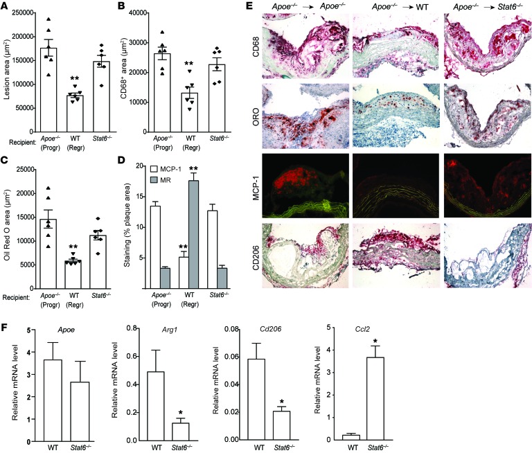
Figure 6. Lack of STAT6 in recipient mice prevents M2 macrophage enrichment of plaques and impairs atherosclerosis regression.
Analysis of aortic arch plaques from mice 3 days after transplantation into Apoe–/– (Progr; n = 6), WT (Regr; n = 6), or Stat6–/– (n = 6) mice for (A) lesion area, (B) immunohistochemical staining for the macrophage marker CD68, (C) Oil red O staining for neutral lipid, and (D) immunohistochemical staining for the M1 macrophage marker MCP-1 and the M2 macrophage marker MR (CD206); **P < 0.001 compared with Apoe–/– progression group using 1-way ANOVA with Dunnett’s multiple comparisons testing. (E) Representative images of aortic plaques stained for CD68, Oil red O (ORO), MCP-1, and CD206, imaged at ×40 magnification. (F) qRT-PCR analysis of mRNA expression of newly recruited monocyte-derived macrophage marker (Apoe), CD206 (M2) (Arg1 and Cd206), and M1 (Ccl2) macrophage markers in CD68+ cells laser captured from the aortic arch plaques 3 days after transplant into WT and Stat6–/– recipient mice (n = 4–7 per group). *P < 0.05 using unpaired t testing.

