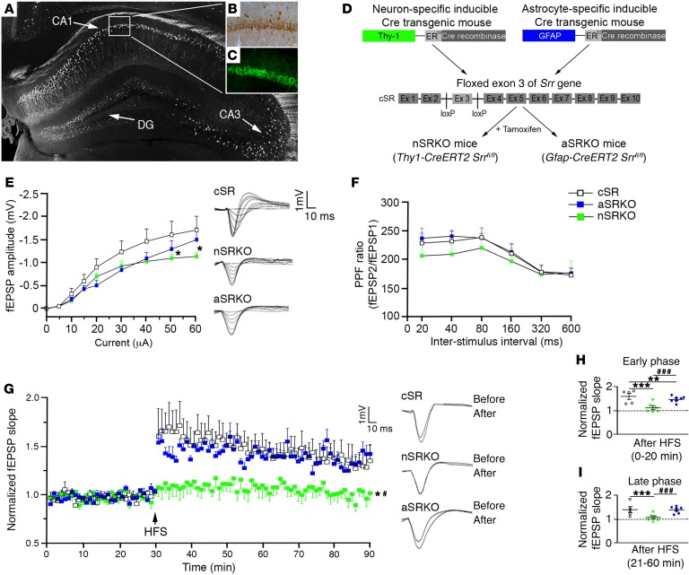
Figure 1. Neuronal d-serine is important for synaptic potentiation.
d-serine (B) and SRR (C) immunoreactivity was observed in the CA1 pyramidal cell layer as outlined in a Thy-1–GFP–labeled hippocampus (A). Scale bar: 25 μm. (D) Schematic flow chart of the nSRKO mouse and the aSRKO mouse. (E) I/O curve showed reduced fEPSP amplitude for nSRKO mice at high intensities. Individual traces shown. (F) PPF showed no significant differences between genotypes. (G) LTP was reduced in nSRKO mice as compared with WT and aSRKO mice. Individual traces shown. (H) Histograph of LTP early phase showed reductions in nSRKO mice. (I) Histograph of LTP late phase showed reductions in nSRKO mice. Data represent mean ± SEM. (E–I) n = 5–8/group. (E–G) Two-way RM ANOVA. (H, I) One-way ANOVA. *P < 0.05; **P < 0.01; ***P < 0.001, compared with cSR control mice. #P < 0.05; ###P < 0.001, compared with nSRKO mice.

