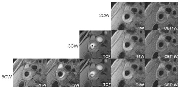
FIGURE 4.
Images are from the right internal carotid artery of an asymptomatic 62-year-old male patient. A fibrous cap rupture was detected after the addition of TOF to the black blood contrast weightings (2CW) because of the presence of jux-taluminal hemorrhage (arrow). No evidence of fibrous cap rupture was found on black blood images alone.

