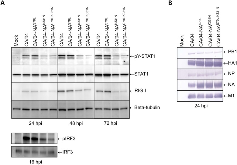
Fig 7.
(A) Comparison of the levels of tyrosine-phosphorylated STAT1, RIG-I, and IRF3 phosphorylation in influenza-infected Calu-3 cells. Cells were infected with the mutant viruses (MOI = 1) and incubated for 16, 24, 48 or 72 h. At the specified time points, whole cell lysates were prepared and the levels of STAT1 activation, RIG-I expression, and IRF3 phosphorylation were measured by Western blot analysis. Total cellular STAT1 and beta-tubulin protein levels were analyzed to control for equal loading. hpi—hours post-infection. (B) Comparison of the PB1, HA1, NA, NP, and M1 levels in influenza-infected Calu-3 cells. Calu-3 cells were infected with the mutant viruses (MOI = 1) and incubated for 24 h. Whole cell lysates were prepared and the levels of viral proteins were measured by Western blot analysis. hpi—hours post-infection.

