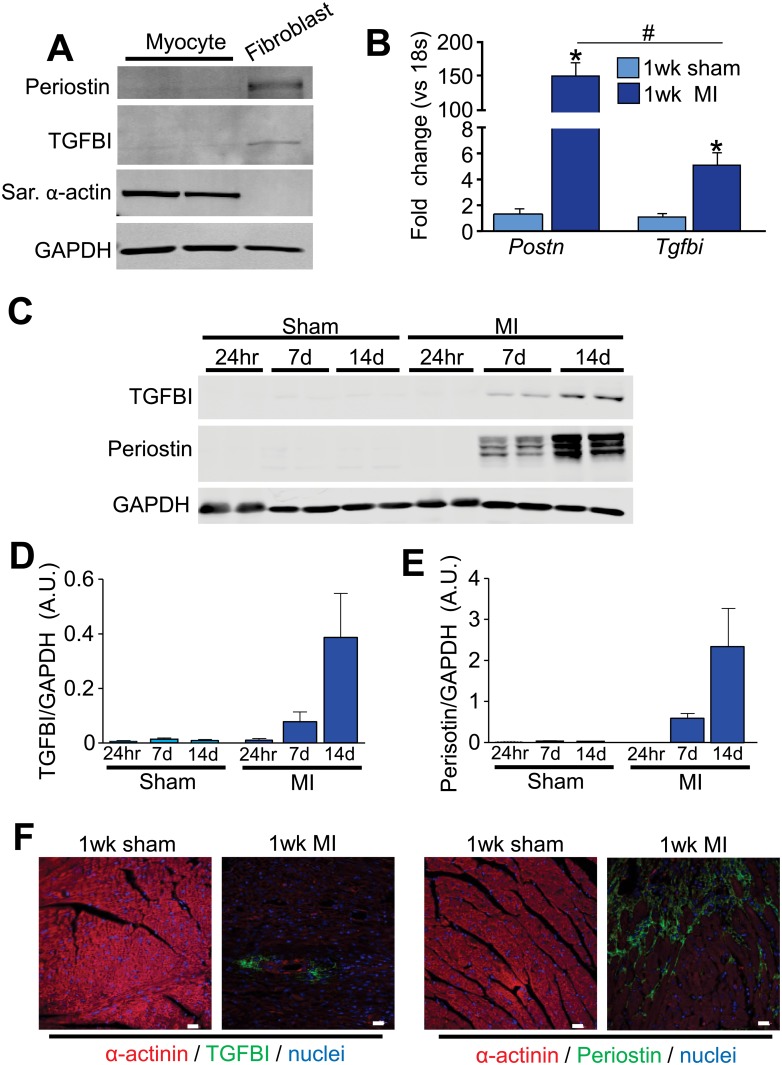
Fig 1. TGFBI and periostin are induced in the heart after injury.
(A) Western blot analysis of periostin and TGFBI in isolated adult cardiomyocytes and fibroblasts. Sarcomeric α-actin was used as a control for cardiomyocyte purity and GAPDH was used as a loading control. (B) Quantitative real time PCR for Postn and Tgfbi from 1 week sham or MI-operated hearts. mRNA levels were normalized to 18s ribosomal RNA. *p<0.05 for both genes in MI-operated animals compared to sham animals using an unpaired Student’s T-test. n = 3 animals. (C) Western blot analysis for periostin and TGFBI in the infarcted areas isolated from hearts 24 hours, 7 and 14 days after MI surgery. GAPDH was used as a loading control. Each lane corresponds to protein from one mouse. (D and E) Quantification of TGFBI (D) and periostin (E) protein levels from the conditions shown in panel C except that 3 hearts were analyzed in total each. (F) Immunohistochemistry for the indicated markers/proteins on sham or MI-operated hearts after 1 week. Images were taken at 400x magnification. Scale = 20 μm.

