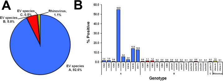
Fig 5. Distribution of EV species and types found in 817 EV-positive HFMD samples.
(A) Pie chart of EV-A to -C and rhinovirus found in the fecal samples of HFMD patients. (B) Genotypes of EV and their percentages (denoted by numbers above the bar graphs). Blue, EV-A; red, EV-B; green, EV-C; yellow, rhinovirus.

