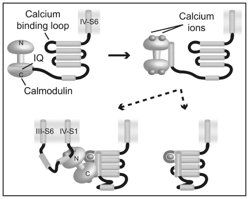
Figure 3.

Model for the action of calcium in the hH1 C-terminus. At basal levels of calcium in the cell, calmodulin (yellow dumbbell) is localized to the IQ motif (light blue cylinder), bound via the C-lobe (top left). As calcium levels rise, calmodulin binds calcium (top right), which weakens and alters the CaM/IQ interaction. The consequent release of one or both CaM domains enables the IQ motif to interact with the EF-hand core (bottom panels), raising its calcium affinity by three orders of magnitude which in turn leads to calcium binding in that site.
