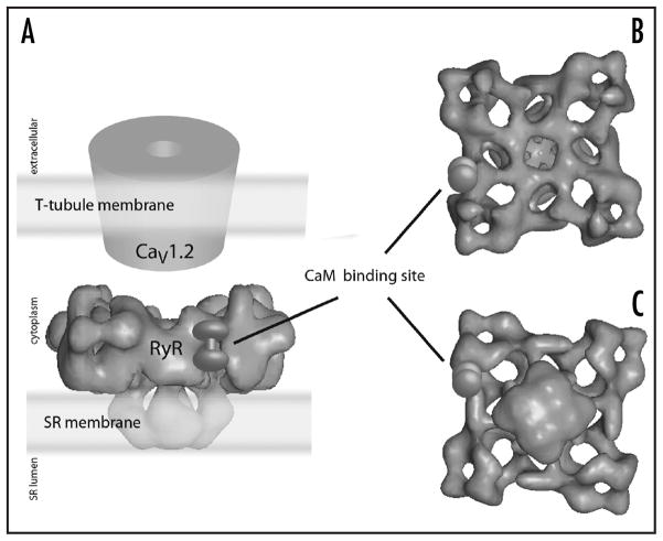
Figure 5.
Three-dimensional reconstruction of the structure of the ryanodine receptor (RyR) tetramer by high-resolution electron microscopy. (A) shows a side view and emphasizes the proximity of RyR to L-type calcium channels in the T-tubule system. (B) shows a view from the cytoplasmic perspective, and (C) shows a view from the SR lumen perspective. The location of calmodulin binding on a single subunit of the tetramer is indicated. Figure adapted with permission from reference 45.

