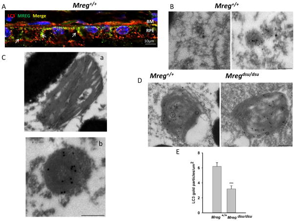
Fig. 7.
LC3 associates with phagosomes and MREG in epithelial cells a MREG and LC3 codistribute in Mreg+/+ (C57Bl6/J) RPE. Eyecups prepared from Mreg+/+ mice (6 months old, 6 h after light onset) were fixed and stained with anti-MREG (mAb, Abnova), shown in green and anti-LC3 rabbit polyclonal (Cell Signaling), shown in red. Pearson’s coefficient 0.74. RPE Retinal Pigment Epithelium; BM Bruch’s Membrane. Scale bar=10 μm. b LC3 and MREG are associated with intracellular vesicles. Scale bar is 100 nm. c LC3 localizes to disk membrane containing structures in mouse RPE. Retinal sections were labeled with anti-MREG mAb165 and anti-LC3 mAb (Abcam) conjugated to gold particles (large-MREG) and (small-LC3). Scale bar is 250 nm. d LC3 localizes to opsin-positive phagosomes. Retinas from C57Bl6/J mice (6 month old, 6 h after light onset) were prepared by embedment in L.R. white resin. Sections were labeled with anti-opsin mAb 4D2 and anti-LC3 mAb (Abcam) conjugated to gold particles. Large particles (opsin) and small particles (LC3). Scale bar is 250 nm. e Phagosomes in Mreg +/+ and Mregdsu/dsu RPE were identified based on RHO ab labeling. The LC3b immunogold labeling was quantified in RHO-positive phagosomes and expressed as gold particles per micrometer squared of phagosome section. (Panel d is a representative image). p<0.01

