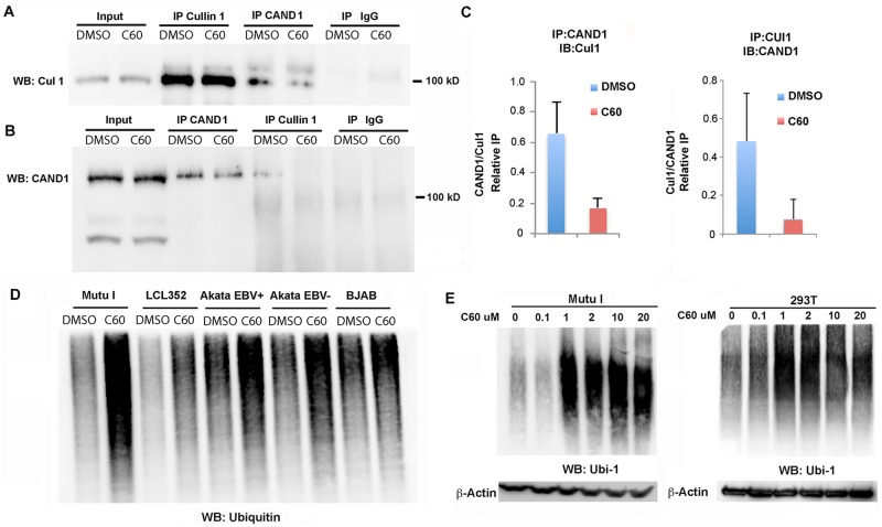
Fig 5. C60 disrupts CAND1-Cullin1 interaction and increases global ubiquitylation.
(A-C) Extracts from Mutu I cells treated with DMSO or C60 for 48 hrs were subject to IP with antibody to Cullin 1, CAND1, or IgG and analyzed by Western blot for Cullin1 (A) or CAND1 (B). Input (10%) is shown in first two lanes. (C) Quantitation of three independent coIP experiments as represented in panels A and B. (D) Mutu I, LCL352, Akata EBV+, Akata EBV-, and BJAB cells were treated with DMSO or C60 for 48 hrs, and then assayed by Western blot with antibody to Ubiquitin 1 (Ubi-1). (E) Dose dependent activation of global ubiquitylation in Mutu I (left) and EBV-negative 283T (right) cells. Total cell extracts were treated with indicated concentration of C60 for 48 hrs. Actin loading control is shown below.

