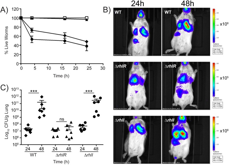
Fig 6. The P. aeruginosa ΔrhlI mutant is virulent and the ΔrhlR mutant is avirulent in animal infection models.
A) C. elegans were applied to lawns of E. coli OP50 (open squares), WT P. aeruginosa PA14 (closed diamonds), ΔrhlR mutant (closed triangles), and ΔrhlI mutant (closed circles). Error bars represent SEM of three independent replicates. B) Real-time monitoring of WT P. aeruginosa PA14 P1-lux and isogenic mutants in the acute pneumonia model. BALB/c mice infected with WT, ΔrhlR, and ΔrhlI strains were imaged at 24 and 48 h using an IVIS CCD camera following intratracheal infection. Imaging was performed from the ventral side of representative mice while the animals were under isoflourane anesthesia. The color bars indicate the intensity of the bioluminescence output, with red and blue denoting the high and low signals, respectively. Note that the color scales on the various mouse bioluminescence imaging panels are not the same. (C) Bacterial load in lung homogenates of mice infected intratracheally with WT P. aeruginosa PA14, and the ΔrhlR and ΔrhlI mutants. Each symbol represents a single mouse. The data are pooled from two independent experiments. Data were analyzed using the Mann-Whitney U test. *** P <0.001 and ns means not significant.

