FIGURE 2:
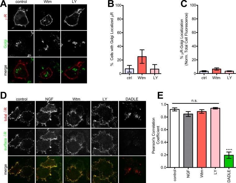
PI3K inhibition does not induce intracellular retention of μR. (A) Representative images (of three independent experiments) of PC12 cells expressing FLAG-μR as a control do not show retention after PI3K inhibition. (B) Quantitation of percentage of cells showing μR retention after PI3K inhibition (>100 cells each; mean ± SEM; not significant [p > 0.05] by two-sided t test vs. control). (C) Quantitation of percentage of total μR fluorescence that overlaps with the Golgi after PI3K inhibition (>100 cells each; mean ± SEM; not significant [p > 0.05] by two-sided t test vs. control). (D) Representative images (of three independent experiments) for δR endocytosis estimated by selectively labeling the surface vs. total pool of δR as described in Materials and Methods. PI3K inhibition did not cause endocytosis. The δR agonist DADLE was used as a control for endocytosis. (E) Pearson’s r of colocalization of the primary and secondary antibodies. High correlation denotes minimal endocytosis. DADLE significantly reduced the correlation, consistent with endocytosis (three representative fields; mean ± SEM; ****p < 0.0001 by two-sided t test vs. control).

