. 2017 Jul 10;6:e26980. doi: 10.7554/eLife.26980

Figure 3. The synthetic lethal interaction between STAG1 and STAG2 is manifested in disease-relevant bladder cancer and Ewing sarcoma cell lines.

(A) The indicated bladder cancer cell lines were analyzed for STAG2 expression by immunoblotting. (B) The indicated bladder cancer cell lines were transfected with NTC, STAG1 and PLK1 siRNA duplexes. Viability was determined 7 or 10 days after transfection and normalized to the viability of NTC siRNA transfected cells (n = 2 independent experiments with 5 biological repeats each, error bars denote standard deviation). (C) STAG2 wild-type UM-UC-5 and STAG2 mutated UM-UC-3 cells were transfected with NTC, STAG1 and SGOL1 siRNA duplexes. Colony formation was analyzed 7 days after transfection by crystal violet staining. (D) 72 hr after siRNA transfection into UM-UC-5 and UM-UC-3 cells, Giemsa-stained chromosome spreads were prepared and analyzed for sister chromatid cohesion phenotypes (n = 100 spreads, error bars denote standard deviation of two independently analyzed slides). (E) The indicated Ewing sarcoma cell lines were analyzed for STAG2 protein expression by immunoblotting. (F) The indicated Ewing sarcoma cell lines were transfected with NTC, STAG1 and SGOL1 siRNA duplexes. Viability was measured 6 days after transfection and normalized to the viability of NTC siRNA transfected cells (n = 3 independent experiments with 3 biological replicates each, error bars denote standard deviation). (G) STAG2 mutated UM-UC-3 cells were transduced with a lentivirus encoding a FLAG-STAG2 transgene. Stably selected cell pools were subsequently transfected with NTC, STAG1 or SGOL1 siRNA duplexes. Viability was measured 7 days after transfection and normalized to the viability of NTC siRNA transfected cells (n = 4 biological replicates, error bars denote standard deviation).

DOI: http://dx.doi.org/10.7554/eLife.26980.012

Figure 3.

Figure 3—figure supplement 1. Patient-derived STAG2 mutations cause STAG1 dependency in engineered isogenic HCT 116 cells.

Figure 3—figure supplement 1.

HCT 116 cell lines engineered to harbor the indicated deleterious patient-derived STAG2 mutations were transfected with NTC, STAG1 and SGOL1 siRNA duplexes. (A) Protein extracts were prepared 48 hr after transfection and analyzed by immunoblotting. (B) Cell viability was measured 7 days after siRNA transfection and plotted normalized to the viability of NTC-transfected cells (n = 4 independent experiments with 5 biological repeats each, error bars denote standard deviation). (C) Sister chromatid cohesion phenotypes were analyzed in Giemsa-stained mitotic chromosome spreads that were prepared 48 hr after siRNA transfection (n = 100 chromosome spreads, error bars denote standard deviation between two independently analyzed slides).
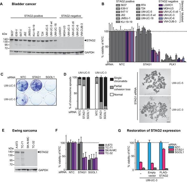
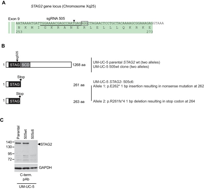
Figure 3—figure supplement 2. Characterization of CRISPR/Cas9-generated STAG2 knockout in UM-UC-5 bladder cancer cell line.

Figure 3—figure supplement 2.

(A) sgRNA 505 co-expressed with Cas9 from an all-in-one plasmid was used to generate deleterious frameshift insertions or deletion mutations (indels) in STAG2 coding exons. The cognate sgRNA target sequence is underlined. Associated protospacer adjacent motifs (PAMs) are marked by boxes. The predicted Cas9 cleavage site is marked by a triangle. (B) CRISPR/Cas9-generated STAG2 indels in the UM-UC-5 cell clone STAG2- 505c6 as detected by Sanger sequencing. STAG2 is located on the X chromosome. Therefore, two alleles had to be inactivated in UM-UC-5 (female) cells. (C) Protein lysates prepared from the indicated parental and mutant UM-UC-5 clones were analyzed for STAG2 protein expression by immunoblotting.

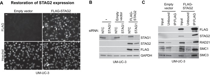
Figure 3—figure supplement 3. Restoration of STAG2 protein expression in UM-UC-3 bladder cancer cells.

Figure 3—figure supplement 3.

STAG2-deficient UM-UC-3 cells were transduced with lentiviral particles encoding no transgene (empty vector) or a 3xFLAG-tagged STAG2 transgene (FLAG-STAG2). Stable selected cell pools were used for further analysis. (A) Expression and nuclear localization of the transgenic FLAG-STAG2 protein was assayed by immunofluorescence microscopy. (B) Non-transduced UM-UC-3 cells, UM-UC-3 cells transduced with an empty vector and UM-UC-3 cells transduced with a FLAG-STAG2 transgene were transfected with NTC or STAG1 siRNA duplexes. Protein lysates prepared 72 hr after siRNA transfection were analyzed by immunoblotting. (C) Protein extracts prepared from UM-UC-3 cells expressing no transgene (empty vector) or a FLAG-STAG2 transgene were subjected to anti-FLAG immunoprecipitation. The input, unbound and precipitated fractions were analyzed by immunoblotting. Co-precipitation of cohesin subunits was only detected in FLAG-STAG2 expressing cells indicating successful incorporation of the transgenic STAG2 protein into the cohesin complex.