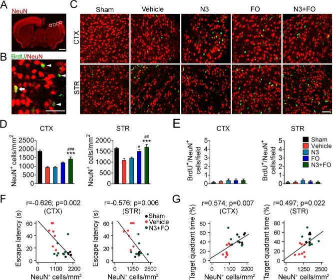
Figure 3.
Post-TBI n-3 PUFA treatment attenuates cortical and striatal neuronal death. (A) A representative image of one coronal brain section showing NeuN immunofluorescence at 35 days after traumatic brain injury (TBI). Boxes demonstrate the ipsilateral perilesion areas in the cortex (CTX) and striatum (STR) where images in (C) were taken. Scale bar: 1 mm. (B) A representative image from the perilesion cortex showing typical double-label immunofluorescence of 5-bromo-2′-deoxyuridine (BrdU) and NeuN. Arrow: BrdU+/NeuN+ cell (yellow). Arrowheads: BrdU+/NeuN− cells (green). Scare bar: 50 μm. (C) Representative images of BrdU (green) and NeuN (red) immunofluorescence in the perilesion cortex and striatum at 35 days after TBI. Scale bar: 50 μm. (D, E) Quantification of total viable neurons (D) and newly generated mature neurons (E) in the perilesion cortex and striatum. *p ≤ 0.05, ***p ≤ 0.001 versus vehicle; ##p ≤ 0.01, ###p ≤ 0.001 versus N3 (fatty acid injection) by one-way analysis of variance (ANOVA). (F, G) Pearson correlation between the mice's spatial learning (F) and memory (G) performance in the Morris water maze and the numbers of total viable neurons in the cortex and striatum. n = 6 mice for sham group; n = 8 mice for vehicle, N3, and fish oil dietary supplementation (FO) groups; n = 7 mice for N3 + FO group.

