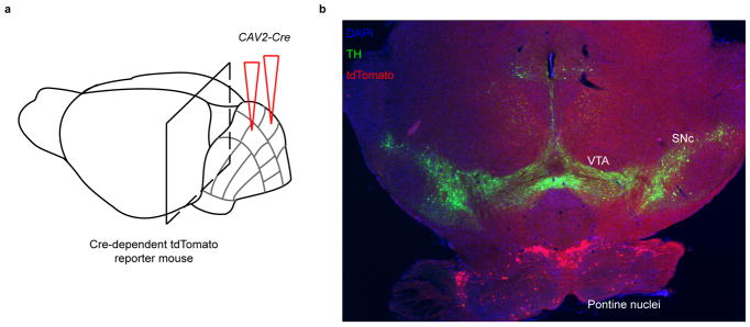
Extended Data Fig. 10. Granule cell reward responses unlikely result from a direct midbrain dopaminergic projection to the cerebellar cortex.
Previous literature on the topic of dopamine in the cerebellum has been controversial, with some anatomical tracing studies suggesting a projection to cerebellar cortex from ventral tegmental area (VTA)39,40, while others failed to find such a projection41. Some studies identified the presence of dopamine in the cerebellar cortex directly42–44, yet a major confound arises due to the large noradrenergic projection to the cerebellum from the locus coeruleus, as dopamine is a precursor to norepinephrine45. To determine whether our widespread reward-related signals were likely to be driven by a direct dopaminergic projection, we traced the inputs to the cerebellar cortex using viral methods. a, Schematic. We injected CAV2-cre, cre recombinase expressed from canine adenovirus-2 known to robustly infect axons and their terminals in many neuronal types46 including dopaminergic neurons specifically47,48, into the cerebellar cortex of a highly sensitive cre-reporter Ai14 transgenic mouse. Thus any neuron in a region presynaptic to the cerebellar injection site infected by CAV2 will express tdTomato. We injected either the vermis of Lobule VI (n =3 mice) or for comparison also the hemisphere lobule crus I (1 mouse). b, We stained serial coronal brain sections for tyrosine hydroxylase (TH, a marker for dopaminergic neurons) and examined the distribution of input cells in the midbrain. In all 4 mice examined (sixty-four 40- or 60-micron sections encompassing all midbrain dopamine neurons), we did not find any VTA or substantia nigra pars compacta (SNc) dopamine neurons projecting to the cerebellar cortex. As a positive control, we noted that all mice exhibited robust tdTomato expression in known inputs to the cerebellum such as the pontine nuclei shown above. To exclude the unlikely possibility that putative VTA dopamine neurons that project to the cerebellum cannot take up CAV2 efficiently, we also performed an experiment where we injected AAVretro-EF1a-FLPo, a virus that robustly infects axonal terminals49, into cerebellar lobule VI of a mouse that expresses FLP-dependent tdTomato, and again did not find tdTomato+ neurons in the VTA or SNc, but abundant tdTomato+ neurons in pontine nuclei (data not shown). Thus if a direct midbrain dopaminergic projection to the cerebellum exists, it must be very sparse, and therefore unlikely to drive the very large and widespread reward-related signals in our granule cell imaging data.

