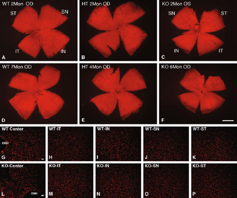
Fig. 2.
Morphometric analysis of PNA-stained cone sheaths. a–f Representative whole mounts stained with PNA from 2 to 7 months. Uniform distribution of cone sheaths was observed in WT, HT, and KO mice retina. Scale bar in F, 1 mm. (g–p) Increased magnification of representative regions of the 2-month retina of (g–k) WT and (l–p) KO in each of five regions: Center, central including optic nerve head (ONH); IT inferior temporal; IN inferior nasal; SN superior nasal; ST superior temporal. Scale bars in G, L, 20 µm

