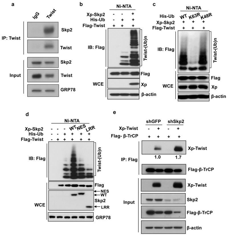
Figure 4. Skp2 interacts with and promotes non-degradative ubiquitination of Twist.
(a) PC3 cells were used for immunoprecipitation (IP) with Twist antibody followed by western blot analysis. (b) In vivo ubiquitination assay in 293T cells transfected with Xpress (Xp) tagged Skp2, Histidine-tagged ubiquitin (His-Ub) and Flag-tagged Twist. Ni-NTA indicates nickel nitrilotriacetic acid bead precipitate; WCE indicates whole cell extract. (c) In vivo ubiquitination assay in 293T cells transfected with various plasmids. (d) In vivo ubiquitination assay in 293T cells transfected with Xp-Skp2 and Flag-Twist along with His-Ub WT, His-Ub K63R or His-Ub K48R constructs. (e) 293 cells with GFP or Skp2 knockdown transfected with Flag-tagged β-TrCP and Xp-tagged Twist were subjected to IP after 48 hours with Flag antibody followed by western blot analysis. The relative intensities of immunoprecipitated Twist were quantified with Image J software and normalized with Twist in the input.

